See the Heart Like Never Before: Transparent 3D Models for Surgical Precision
6/5/20252 min read


Introduction
Understanding the heart’s complex anatomy is critical for both surgeons and medical students. Traditional 2D imaging or textbooks can only take you so far. With intricate chambers, valves, and conduction pathways, visualizing spatial relationships accurately is challenging.
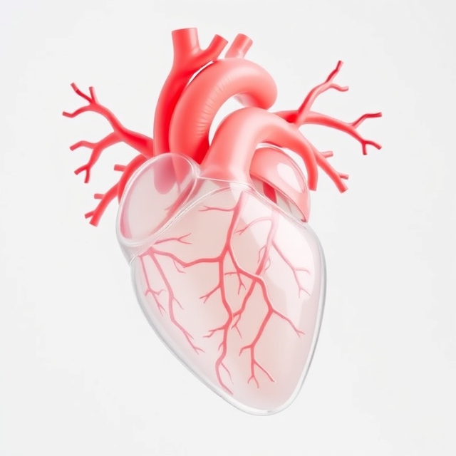
Transparent 3D printed heart models transform the learning and planning experience. By combining tactile feedback with precise anatomical accuracy, these models make internal structures visible. Surgeons can plan, practice, and teach with confidence.
The Challenge of Cardiac Anatomy
The heart is a compact organ with overlapping structures:
Four chambers (atria and ventricles)
Valves controlling directional blood flow
Coronary arteries and veins supplying the myocardium
Conduction pathways that dictate rhythm
Even experienced surgeons often rely heavily on imaging for orientation. Translating 2D scans into 3D spatial understanding requires experience and can lead to errors during procedures.
How Transparent Heart Models Help
1. Pre-Operative Planning
Patient-specific heart models allow surgeons to visualize anomalies, complex pathologies, or unusual vessel arrangements before surgery. Practicing on a model reduces surprises and improves precision.
2. Education and Training
Medical students and residents benefit from hands-on experience. Seeing the heart’s internal structures in 3D helps solidify spatial understanding that textbooks or screens cannot provide.
3. Visualization of Conduction Pathways
Transparent models make it easier to study pathways for procedures like LBBA pacing. Clinicians can anticipate challenges and refine their technique.
4. Patient Communication
A physical 3D heart model makes complex conditions more understandable. Patients and families can see exactly what the procedure involves, improving trust and informed consent.
Why Transparency Matters
Unlike opaque models, transparent 3D prints let you see internal structures without disassembly. Surgeons can observe:
Valve function
Blood flow paths
Chamber orientation
Conduction routes
This level of detail is valuable for teaching, planning surgeries, and presenting complex cases.
From Scan to Model
Our process turns CT or MRI data into a high-precision 3D model:
Data Processing: Extracting anatomical structures from scans
STL Preparation: Ensuring accuracy for printing
3D Printing: Using transparent resin to maintain clarity and detail
Finishing: Polishing for smooth, fully transparent visualization
The result is a model that is both educational and functional. It can be used repeatedly for planning, teaching, or simulation.
Conclusion
Transparent 3D heart models are more than educational tools. They are a bridge between imaging, surgery, and understanding. Surgeons can plan with confidence, students can learn more effectively, and patients can visualize their care.
Contact us today to see how a transparent heart model can support your surgical planning, training, or patient education. Our team will work with you to create a model tailored to your needs.
info@clearsimulations.com
Precision
Custom 3D printed anatomical models for education.
info@clearsimulations.com
(+1) 813 455 9388
© 2024. All rights reserved.
Contact
Subscribe
Subscribe for exclusive deals, updates, and the latest product launches!
