The Advantages of Transparent Anatomical Models in Education, Training, and Surgical Planning
9/7/20243 min read
The Advantages of Transparent Anatomical Models in Education, Training, and Surgical Planning
Transparent anatomical models are becoming indispensable tools in medical and dental fields, offering clear benefits for education, training, and surgical planning. Made possible through advancements in 3D printing and materials technology, these models allow both students and practitioners to observe internal structures and complexities in a way that traditional models cannot. Here are some of the major advantages of using transparent anatomical models in clinical and educational settings.
1. Enhanced Visualization of Internal Structures
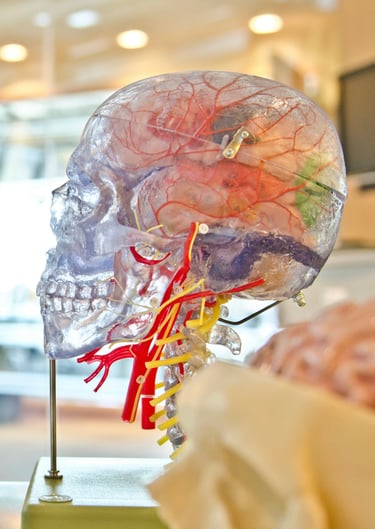
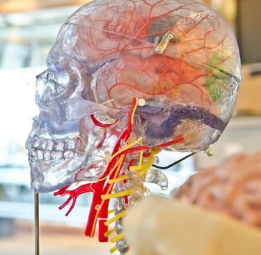
Transparent models offer a clear view of internal anatomy without the need to disassemble or slice through the model. This is particularly beneficial for areas with intricate structures, such as the oral cavity, where nerves, blood vessels, and other critical structures are in close proximity.
For surgical planning, these models enable surgeons to see exactly where they need to operate and what internal structures to avoid. Dentists planning complex root canals, for example, can study a model that accurately reflects the intricate root pathways and inner anatomy of the teeth. This level of detail can prevent mistakes and enhance the precision of surgery, leading to better patient outcomes.
2. Improved Learning and Training for Students and Trainees
For students and new practitioners, transparent models provide a hands-on, three-dimensional understanding of anatomy, which is essential for mastering complex medical and dental procedures. Unlike textbooks or two-dimensional images, transparent models allow students to view all layers of an anatomical structure in one cohesive model. This approach enables deeper comprehension, as learners can observe spatial relationships and anatomical nuances that are otherwise difficult to grasp.
In surgical training, transparent models help trainees build confidence by simulating real-life scenarios. For example, dental students can practice procedures on a transparent model, visualizing root canals and pulp chambers as they would in an actual patient. This direct experience with realistic anatomy leads to a smoother transition from training to real patient care.
3. Better Communication with Patients
Transparent models are also powerful tools for patient education, helping individuals understand their diagnoses and treatment plans. Showing patients a transparent model of their affected area makes it easier for them to visualize the problem and understand why a particular surgical approach is necessary. For example, a dentist can use a transparent model to demonstrate the complexity of an impacted tooth or the placement of an implant, allowing the patient to see how their oral anatomy will be impacted by the procedure.
This level of visualization not only helps patients feel more informed but can also alleviate anxiety about the procedure, as they gain a clearer understanding of what will happen during surgery. By fostering this level of clarity and transparency (literally), dental professionals can improve patient satisfaction and build trust.
4. Streamlined Surgical Planning and Reduced Operating Time
For surgeons, detailed pre-operative planning is critical to ensure successful outcomes. Transparent models provide a roadmap to understand the full scope of anatomical structures, identifying potential complications before the procedure begins. Being able to see through layers, understand depths, and determine the safest approach ahead of time helps surgeons make quicker, more informed decisions.
With this clear understanding, surgeons can anticipate and avoid potential obstacles, such as nerve damage or interference with blood vessels. As a result, operating times can be significantly reduced, improving efficiency, reducing patient time under anesthesia, and lowering the risk of complications.
5. Cost-Effectiveness and Sustainability in Medical Training
The use of transparent 3D-printed models in training offers a reusable, customizable, and cost-effective alternative to traditional cadaveric dissections or animal models, which can be resource-intensive and expensive. By creating transparent models based on digital scans, educational institutions and medical practices can produce realistic, patient-specific models tailored for various training purposes. These models can be reused for multiple training sessions, making them a more sustainable solution for medical education and training programs.
Conclusion
Transparent anatomical models have redefined the standards for medical and dental education, training, and surgical planning. They offer unparalleled visualization of internal structures, enabling better understanding, enhanced surgical precision, and improved patient communication. As 3D printing technology continues to advance, the future of transparent anatomical models looks even more promising, potentially making these tools accessible to more practices, students, and patients around the world.
Precision
Custom 3D printed anatomical models for education.
info@clearsimulations.com
© 2026. All rights reserved.
Contact
Subscribe
Subscribe for exclusive deals, updates, and the latest product launches!